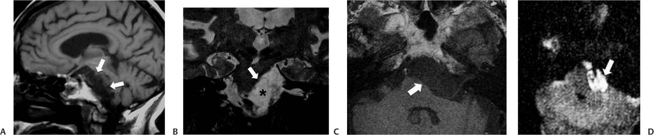
Case 24 A 35-year-old man with progressive left-sided hearing loss. (A) Sagittal T1-weighted image (WI) shows the irregular anterior border of the pons (arrows). (B) Coronal T2WI demonstrates displacement of the pons to the right (arrow). A lesion in the left cerebellopontine angle (CPA) has a slightly higher signal than that of cerebrospinal fluid (CSF; asterisk). (C) Axial T1WI shows a lesion in the left CPA that has a signal like that of CSF and displaces the pons to the right (arrow). (D) Diffusion-WI demonstrates high signal in the mass located in the left CPA (arrow). • Epidermoid cyst:
Clinical Presentation
Imaging Findings
Differential Diagnosis
![]()
Stay updated, free articles. Join our Telegram channel

Full access? Get Clinical Tree


