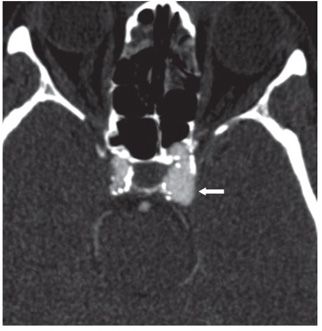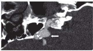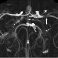

FINDINGS Figure 24-1. Axial NCCT through the sella. There is a left parasellar hyperdense ovoid lesion that occupies and bulges the left cavernous sinus (arrow). Figures 24-2 and 24-3. Axial and sagittal reconstructed views, respectively, from CTA in the same patient. There is an intensely enhancing saccular aneurysm of the left cavernous internal carotid artery (ICA) (arrows).
DIFFERENTIAL DIAGNOSIS Pituitary adenoma, meningioma, cavernous sinus wall hemangioma, metastases, schwannoma, parasellar aneurysm.
DIAGNOSIS Parasellar aneurysm.
DISCUSSION
Stay updated, free articles. Join our Telegram channel

Full access? Get Clinical Tree


