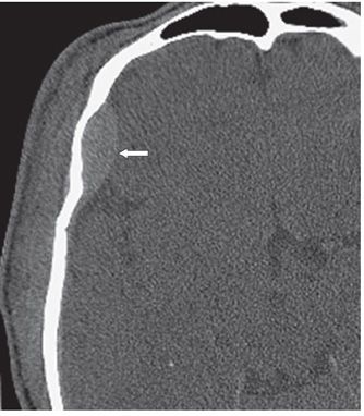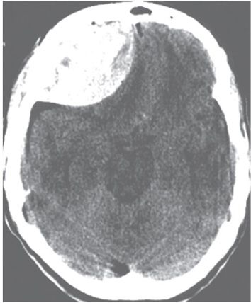

FINDINGS Figure 240-1. Axial NCCT through the frontal lobes. There is a right frontal convexity extraaxial biconvex hyperdensity measuring 1.1 cm in maximum thickness displacing the brain medially from the inner table (arrow). There is overlying subgaleal hematoma and swelling of the scalp. Figure 240-2. Axial NCCT bone algorithm “blood window level” 1.25 mm reconstruction at a level inferior to Figure 240-1. This emphasizes the biconvex nature of the hyperdensity. There is a hairline fracture of the overlying frontal bone (not shown). Figure 240-3. Axial NCCT in a different patient. This demonstrates a very large biconvex mixed density but mostly hyperdense right frontal acute extraaxial hematoma with significant mass effect on the right frontal lobe. The mixed density suggests active bleeding.
DIFFERENTIAL DIAGNOSIS Acute subdural hematoma (ASDH), acute epidural hematoma (AEDH).
DIAGNOSIS Acute epidural hematoma (AEDH).
DISCUSSION
Stay updated, free articles. Join our Telegram channel

Full access? Get Clinical Tree


