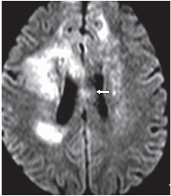
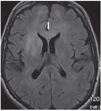
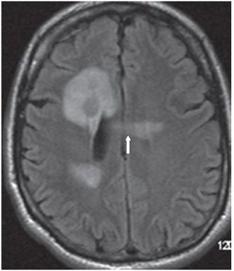
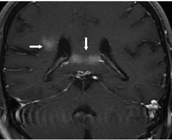
FINDINGS Figures 242-1 and 242-2. Axial DWI through the splenium and body of corpus callosum (CC), respectively. There are smudgy hyperintensities in the splenium (vertical arrow in Figure 242-1) and body of CC (transverse arrow in Figure 242-2). These areas show low ADC. Similar smudgy hyperintensities are present in bilateral periventricular white matter (WM). Figure 242-3. Axial FLAIR through the genu of CC. There is smudgy hyperintensity extending from the right frontal WM to the left through the genu of CC (arrow) the so-called butterfly lesion. Figure 242-4. Axial FLAIR through the body of CC. There is a transverse band of smudgy hyperintensity through the mid body of the CC (vertical arrow). Similar smudgy periventricular hyperintensities are present in the right corona radiata with the right frontal lesion showing an iso/hypointense core. Figure 242-5. Coronal post-contrast T1WI through the splenium. There is a band of contrast enhancement through the splenium (vertical arrow) and in the right corona radiata (transverse arrow). Similarly there is smudgy contrast enhancement across the body of CC (not shown).
DIFFERENTIAL DIAGNOSIS Acute disseminated encephalomyelitis (ADEM), multiple sclerosis (MS), lymphoma, butterfly glioma.
DIAGNOSIS


Stay updated, free articles. Join our Telegram channel

Full access? Get Clinical Tree


