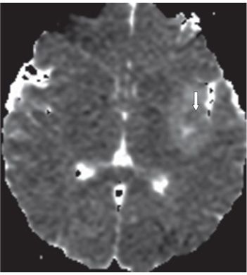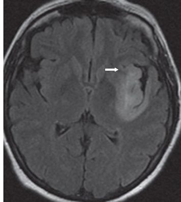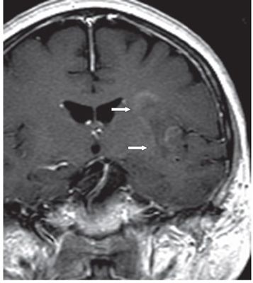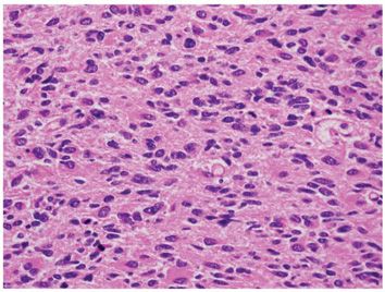


FINDINGS Figures 243-1 and 243-2. Axial DWI and corresponding ADC map through the sylvian region. There is a left insula/subinsula minimal opercula diffuse hyperintensity on DWI with a small focal area of diffusion restriction in the insula on ADC map (arrow). Figure 243-3. Axial FLAIR through the lesion. There is diffuse hyperintensity in the left insula and surrounding region with apparently well-defined margin except anteriorly (arrow). There is no significant mass effect. Figure 243-4. Coronal post-contrast T1WI through the lesion. There is patchy lineal predominant peripheral enhancement (arrows). Figure 243-5. Photomicrograph shows hypercellular tumor with atypical hyperchromatic nuclei and several mitoses.
DIFFERENTIAL DIAGNOSIS Subacute enhancing MCA infarct, encephalitis (especially HSV), progressive multifocal leukoencephalopathy (PML), anaplastic astrocytoma (AA) WHO III, astrocytoma WHO II, changes due to seizure.
DIAGNOSIS Anaplastic astrocytoma (AA).
DISCUSSION
Stay updated, free articles. Join our Telegram channel

Full access? Get Clinical Tree


