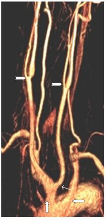
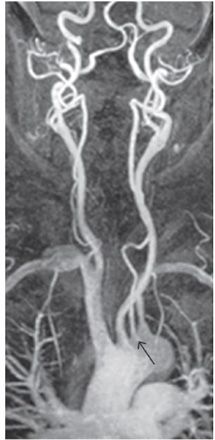
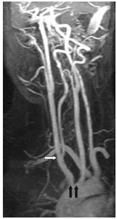
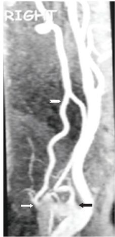
FINDINGS All figures are contrast-enhanced MIP or volume-rendering neck MRA in different projections to best demonstrate the anatomy. Figure 244-1. Normal three-vessel origins from the aortic arch. From right to left are the brachiocephalic trunk, which divides into the right subclavian artery and the right common carotid artery (RCCA), the left common carotid artery (LCCA), and the left subclavian artery. The vertebral arteries (transverse arrows) originate from the subclavian arteries. The common carotid artery (CCA) divides distally into the internal carotid artery (ICA) and the external carotid artery (ECA) at about C4 level. This is the most common aortic arch configuration encountered. Figure 244-2. There is a common origin of the brachiocephalic trunk and the LCCA—the so-called bovine origin (arrow) which is different from the real bovine configuration. This is a common anomaly. The true bovine configuration has a single trunk from the aortic arch that gives rise to all the neck and head vessels. The LCCA (line arrow) arises as a branch of the brachiocephalic trunk in this case. The left subclavian artery has a separate origin (transverse arrow). The chevrons point to the CCA bifurcations. Figure 244-3


Stay updated, free articles. Join our Telegram channel

Full access? Get Clinical Tree


