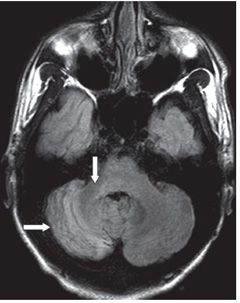
FINDINGS Figure 245-1. Axial FLAIR image through the cerebrum. There is an old left hemispheric watershed infarct (arrows) with left hemispheric atrophy. Figure 245-2. Axial FLAIR through the cerebellum. There is atrophy and smudgy hyperintensity within the right cerebellar hemisphere, right brachium pontis, and pons (arrows).
DIFFERENTIAL DIAGNOSIS Crossed cerebellar atrophy and diaschisis, primary cerebellar infarct, chronic infection/inflammation of the cerebellum.
Stay updated, free articles. Join our Telegram channel

Full access? Get Clinical Tree


