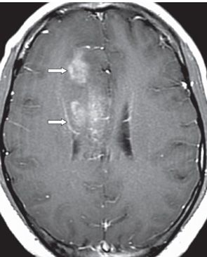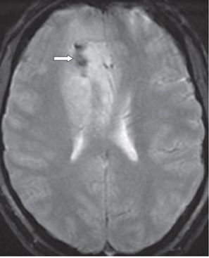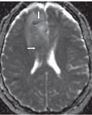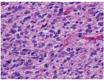


FINDINGS Figure 248-1. Axial FLAIR MRI through the level of the corpus callosum. There is a mildly heterogeneous hyperintense mass in the medial right frontal lobe that is diffusely infiltrating the cortex as well as the adjacent white matter (WM) and corpus callosum. It is effacing the adjacent sulci and the right lateral ventricle with minimal right to left midline shift. Figure 248-2. Axial post-contrast T1WI through the mass. There are multifocal areas of enhancement within the mass (arrows). Figure 248-3. Axial GRE through the mass. There is a small area of hypointensity (arrow) anterolaterally within the mass consistent with calcification or hemorrhage. Figure 248-4. Axial ADC map through the mass. There is relative restricted diffusion (arrows) in the mid and posterior aspect of the mass involving WM with elevated ADC values predominantly anteriorly and at the periphery posterior of the mass involving the GM. Figure 248-5. Photomicrograph shows oligodendroglioma with slight nuclear pleomorphism and atypia and several mitoses (H&E stain).
DIFFERENTIAL DIAGNOSIS Oligodendroglioma, infarct, metastatic disease, infection, status epilepticus, dysembryoplastic neuroepithelial tumor (DNET), ganglioglioma, pleomorphic xanthoastrocytoma (PXA).
DIAGNOSIS
Stay updated, free articles. Join our Telegram channel

Full access? Get Clinical Tree


