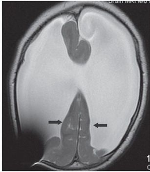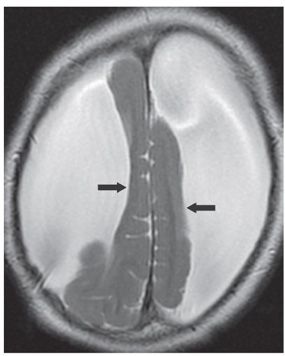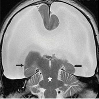


FINDINGS Figure 249-1. Axial T2WI through the thalami. The frontal lobes are absent and replaced by cerebrospinal fluid (CSF) (stars). There is a slit third ventricle between the thalami (transverse arrow). The superior vermis and cerebellum are normal (chevrons). Figures 249-2 and 249-3. Axial T2WI through the expected levels of the lateral ventricles and the centrum semiovale, respectively. Only small parafalcine strips of frontal parietal lobes are present bilaterally (arrows) with the supratentorial space occupied by CSF. The remnants of the frontal lobes display rounded margins. It is impossible to identify convexity brain mantle. Large signal void artifact posteriorly on the right in Figures 249-1 and 249-2 is from ventriculoperitoneal (VP) shunt valve. Figure 249-4
Stay updated, free articles. Join our Telegram channel

Full access? Get Clinical Tree


