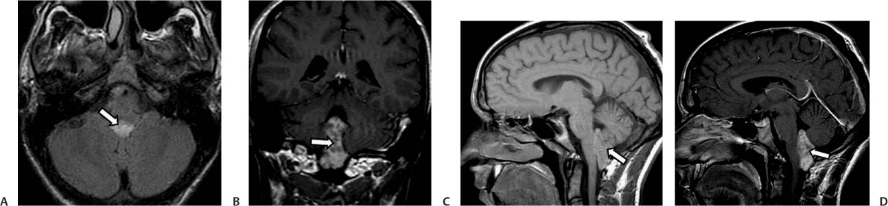
Case 25 A 28-year-old man presenting with ataxia, headache, nausea, and vomiting. (A) Axial fluid-attenuated inversion recovery (FLAIR) image shows a well-defined hyperdense lesion occupying the 4th ventricle (arrow). (B) Coronal T1-weighted image (WI) with contrast shows heterogeneous enhancement of the 4th ventricular mass (arrow). (C) Sagittal T1WI demonstrates an isointense mass (arrow) that occupies the lower aspect of the 4th ventricle. (D) Sagittal T1WI with contrast shows the heterogeneous enhancement of the mass (arrow). • Ependymoma:
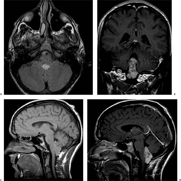
Clinical Presentation
Imaging Findings
Differential Diagnosis
![]()
Stay updated, free articles. Join our Telegram channel

Full access? Get Clinical Tree


