 Clinical Presentation
A 46-year-old woman with Sjögren syndrome.
 Imaging Findings
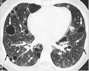
Thoracic computed tomography lung window image demonstrates a thin-walled cyst as well as small nodules (white arrows) and minimal interlobular septal thickening in both lungs (black arrows).
 Differential Diagnosis
• Lymphocytic interstitial pneumonia (LIP): In the clinical setting of a patient with Sjögren syndrome, the presence of small, thin-walled pulmonary cysts is consistent with LIP.
• Pneumocystis jiroveci infection: Cystic changes with thin-walled cysts, nodules, and ground-glass opacities are common findings in Pneumocystis infection.
Stay updated, free articles. Join our Telegram channel

Full access? Get Clinical Tree


