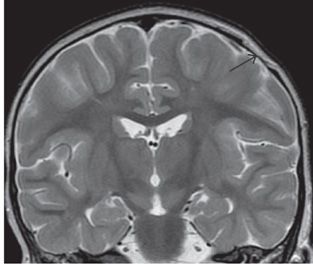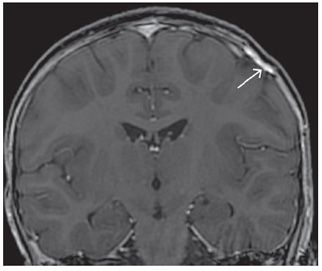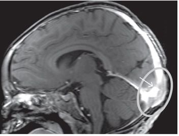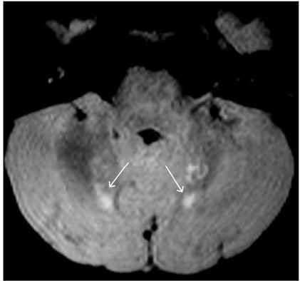


FINDINGS Figure 250-1. Lateral skull X-ray. There is a well-circumscribed, lytic lesion of the parietal calvarium with no marginal sclerosis (anterior arrow). There is a second more geographic lesion near the torcula (posterior arrow). Figure 250-2. Coronal T2WI through the parietal lesion. The lesion shows beveled margins with involvement of the outer table and sparing of the inner table (arrow). Figure 250-3. Coronal post-contrast image shows the bony replacement by mildly enhancing soft tissue (arrow). Figure 250-4. Sagittal post-contrast T1WI demonstrates enhancing soft tissue (circle) with bony destruction at the torcula, (corresponding to the calvarial defect in Figure 250-1), encasing, and narrowing the confluence of venous sinuses (arrow). Figure 250-5. Axial FLAIR through the cerebellum demonstrates T2 hyperintense foci involving the cerebellar dentate nuclei bilaterally (arrows).
DIFFERENTIAL DIAGNOSIS Metastatic disease, infection, epidermoid, dermoid, leptomeningeal cyst, mastoiditis, rhabdomyosarcoma, granulomatous disease, that is, sarcoidosis.
DIAGNOSIS Langerhans cell histiocytosis (LCH) of the bone.
DISCUSSION
Stay updated, free articles. Join our Telegram channel

Full access? Get Clinical Tree


