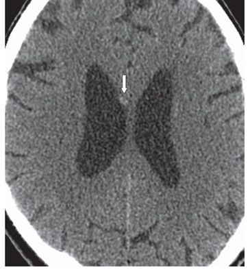
FINDINGS Figure 251-1. Axial NCCT through the body of corpus callosum (CC). There is a focal hyperdensity anteriorly to the right in the body of the CC (arrow). Figure 251-2. Axial NCCT through the same level about 1 month later. There is persistent but smaller hyperdensity in same location.
DIFFERENTIAL DIAGNOSIS Hematoma, calcification.
DIAGNOSIS Corpus callosum hematoma (CCH).
DISCUSSION


Stay updated, free articles. Join our Telegram channel

Full access? Get Clinical Tree


