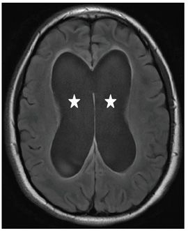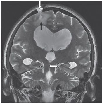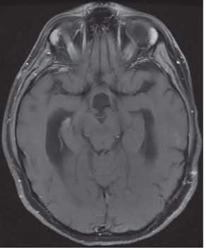


FINDINGS Figure 253-1. Sagittal T1WI through the level of the aqueduct. There is dilation of the third and lateral ventricles (stars). The aqueduct is closed (arrow). The fourth ventricle is normal (chevron). Figure 253-2. Axial FLAIR through the body of the lateral ventricles. There is dilation of the lateral ventricles (stars). There is very thin periventricular hyperintensity suggesting mild transependymal cerebrospinal fluid (CSF) flow. Figure 253-3. Coronal T2WI through the aqueduct. There is dilation of the third and lateral ventricles. The aqueduct is barely visible (transverse arrow). There is a right parasagittal parietal burr hole with underlying encephalomalacia (vertical arrows) from prior intervention. Figure 253-4. Axial post-contrast T1WI through the aqueduct. There is no mass or abnormal contrast enhancement in the brainstem or around the aqueduct.
DIFFERENTIAL DIAGNOSIS Obstructive hydrocephalus, communicating hydrocephalus, volume loss.
DIAGNOSIS Hydrocephalus due to aqueductal stenosis.
DISCUSSION
Stay updated, free articles. Join our Telegram channel

Full access? Get Clinical Tree


