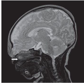
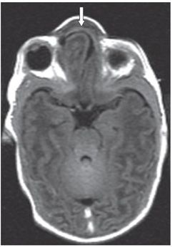
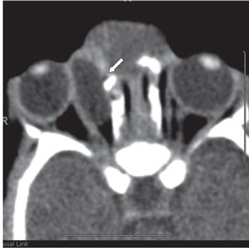
FINDINGS Figure 254-1. Coronal T2WI through the orbits. There is a cystic mass within the right orbit, medial to the globe, and medial rectus muscle (arrow). Figures 254-2 and 254-3. Sagittal T2WI and axial FLAIR, respectively, through the orbits. There is abnormal protrusion of brain parenchyma into the nasofrontal region (arrows). Figure 254-4. Axial CECT through the orbits. There is a thin communication (white arrow) between the orbital cyst and the nasal abnormality.
DIFFERENTIAL DIAGNOSIS Mucocele, dermoid/epidermoid, dacryocystocele, meningocele.
DIAGNOSIS Orbital meningocele with nasofrontal meningoencephalocele.
DISCUSSION


Stay updated, free articles. Join our Telegram channel

Full access? Get Clinical Tree


