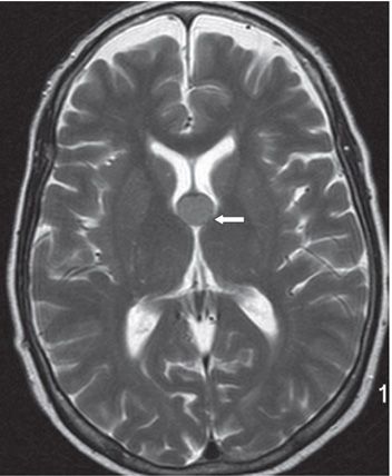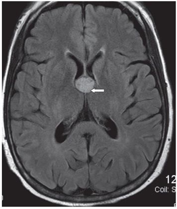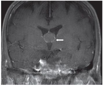


FINDINGS Figure 255-1. Axial NCCT through the frontal horns of the lateral ventricles. There is a round smooth-walled hyperdense 1.2-cm mass at the level of foramina of Monro (arrow). There is no hydrocephalus. Figure 255-2. Axial T2WI through the mass. There is a smooth, round relatively isointense (to gray matter [GM]) mass occupying the region of foramina of Monro (arrow). Figure 255-3. Axial FLAIR. There is a homogeneous hyperintense mass at the foramina of Monro. Figure 255-4. Coronal post-contrast T1WI through the mass. The mass is isointense without contrast enhancement. There is a thin rim of contrast enhancement considered to be within vascular structures or the choroid plexus. Mass hangs down from the septum pellucidum.
DIFFERENTIAL DIAGNOSIS Colloid cyst, choroid plexus papilloma (CPP), choroid plexus carcinoma (CPC), pilocytic astrocytoma, subependymal giant cell astrocytoma (SGCA).
DIAGNOSIS Colloid cyst of the third ventricle.
DISCUSSION
Stay updated, free articles. Join our Telegram channel

Full access? Get Clinical Tree


