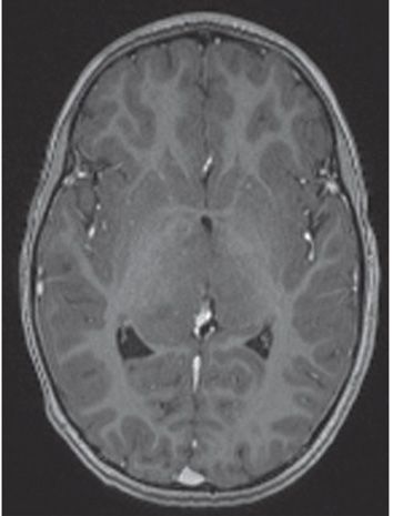
FINDINGS Figure 257-1. Axial FLAIR through the thalami. There is bilateral diffuse enlargement and homogeneous hyperintensity of the thalami. Figure 257-2. Axial T1WI through the thalami. There is non-contrast-enhancing isointensity of the thalami. There is no hydrocephalus.
DIFFERENTIAL DIAGNOSIS Encephalitis, mitochondrial encephalopathy, acute disseminated encephalopathy (rare without white matter involvement) or acute necrotizing encephalopathy of childhood, astrocytoma, germinoma (very rare).
DIAGNOSIS Primary bilateral thalamic astrocytoma.
DISCUSSION
Stay updated, free articles. Join our Telegram channel

Full access? Get Clinical Tree


