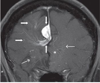
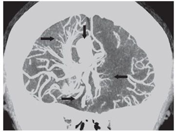
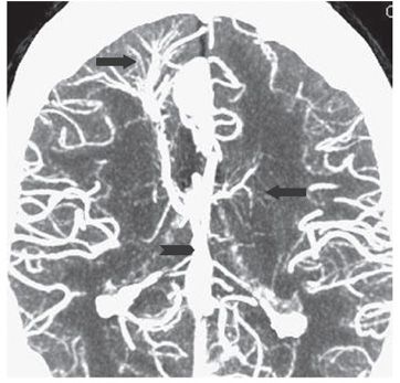
FINDINGS Figure 258-1. Coronal T2WI through the frontal horns. There are prominent right frontal lobe medullary venous signal voids extending from the subcortical region to the right frontal horn (arrows). Figure 258-2. Coronal post-contrast T1WI just anterior to Figure 258-1. There are multiple contrast-enhancing medullary veins in the right frontal corona radiata and centrum semiovale (transverse arrows), the so-called caput medusa draining into a large collector vein (inferior vertical arrow) over the anterior body of the corpus callosum which empties into a large right parafalcine vein (superior vertical arrow). There are also multiple contrast-enhancing venous structures in the bilateral inferior frontal lobes (line arrows) similar to the right frontal corona radiata medullary veins. Figure 258-3. CTA coronal MIP through the frontal lobes. There are multiple bilateral large medullary veins (transverse arrows) draining into the large right parafalcine vein (vertical arrow). Figure 258-4


Stay updated, free articles. Join our Telegram channel

Full access? Get Clinical Tree


