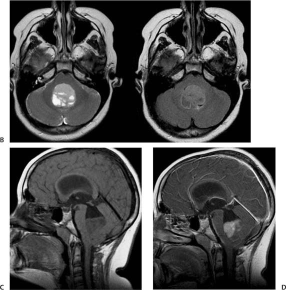
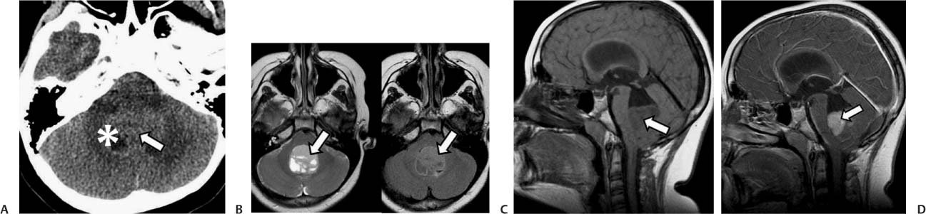
Case 26 A 7-year-old girl with headaches. (A) Computed tomography (CT) scan of the head without contrast shows a slightly hyperdense mass in the area of the 4th ventricle (arrow); there is a cystic part of the lesion (asterisk). (B) Axial T2-weighted and fluid-attenuated inversion recovery (FLAIR) images show a mass centered in the 4th ventricle that has intermediate signal with cystic components (arrows). (C) Sagittal T1-weighted image (WI) without contrast shows the mass (arrow), which is slightly hypointense to the white matter, occupying the lower aspect of the 4th ventricle. Note the dilatation of the entire ventricular system. (D) Sagittal T1WI with contrast shows enhancement of the mass (arrow). • Medulloblastoma:

Clinical Presentation
Further Work-up
Imaging Findings
Differential Diagnosis
![]()
Stay updated, free articles. Join our Telegram channel

Full access? Get Clinical Tree


