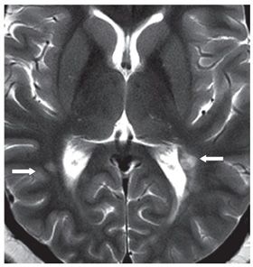
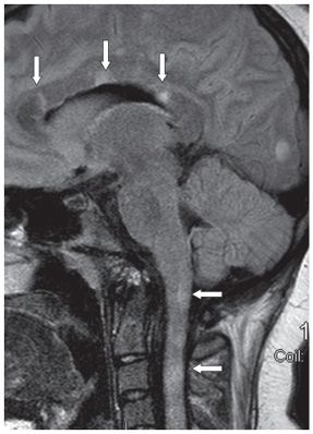
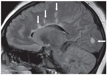
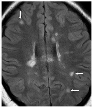
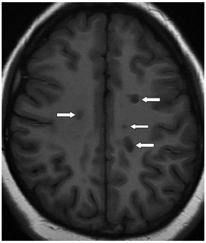
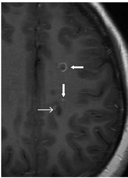
FINDINGS Figure 26-1. Axial MR T2WI through the lower brainstem. There are multifocal brainstem, bilateral brachium pontis, and cerebellar hyperintense lesions (arrows). Figure 26-2. Axial T2WI through the trigones of the lateral ventricles. There are bilateral peritrigonal hyperintense lesions (arrows). Figure 26-3. Sagittal FLAIR through the corpus callosum showing multifocal corpus callosal hyperintense lesions of varying shapes and sizes abutting the callososeptal interface (vertical arrows). There are also multiple cervical spinal cord hyperintense lesions (transverse arrows). Figure 26-4. Parasagittal FLAIR showing multiple ovoid periventricular and deep WM lesions (so-called Dawson fingers, vertical arrows). There is an occipital subcortical lesion (transverse arrow). Figure 26-5. Axial FLAIR through the upper corona radiata showing multiple subcortical lesions (arrows). Multiple periventricular and deep white matter (WM) lesions are also demonstrated. Lesions are of varying shapes and sizes. Figure 26-6. Axial T1WI through the centrum semiovale. There are deep WM multifocal hypointense lesions, the so-called T1-weighted black holes (transverse arrows). Figure 26-7. Axial post-contrast T1WI through the same level. There are multiple contrast-enhancing lesions of the incomplete ring (transverse arrow), punctate (vertical arrow), and a small arc enhancement (line transverse arrow) in the left centrum semiovale.
DIFFERENTIAL DIAGNOSIS Multiple sclerosis (MS), vasculitis, sarcoidosis, Susac syndrome, acute disseminated encephalomyelitis (ADEM).
Stay updated, free articles. Join our Telegram channel

Full access? Get Clinical Tree


