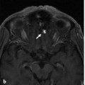
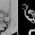
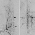
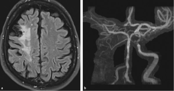
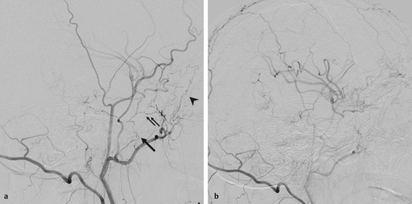
The “Dangerous” Anastomoses II: Petrous and Cavernous Anastomoses A 59-year-old man presented with two right-sided hemodynamic hemispheric ischemic strokes, the last one 10 months before the angiogram. He is a heavy smoker and has a history of peripheral and coronary artery vascular disease. He was being considered for extracranial–intracranial bypass. See ▶ Fig. 26.1, ▶ Fig. 26.2. Fig. 26.1 FLAIR MRI (a) shows infarction in the right frontoparietal lobe with associated volume loss. Small T2 bright spots are observed in the remaining white matter. MRA (b) reveals nonvisualization of the right ICA, likely because of chronic occlusion, and the right A1 segment, which could be related to severe hypoplasia and/or stenosis. The right middle cerebral artery branches are also poorly visualized. Irregularity of the basilar artery and some narrowing of cavernous left ICA are observed, suggesting diffuse underlying atherosclerotic disease. Case is continued in ▶ Fig. 26.2. Fig. 26.2 Cerebral angiography confirmed the right ICA occlusion. There was no A1 segment and minimal leptomeningeal collaterals to the distal middle cerebral artery branches from the PCA branches. Small collaterals through anastomotic channels from the right ECA to the middle cerebral artery were also observed; however, these were insufficient, and there was a delayed venous phase within the middle cerebral artery territory. Right ECA angiogram in lateral view in early (a) and late (b) arterial phases demonstrates collaterals to the ICA from the artery of the foramen ovale (arrow) and the vidian artery (thin double arrows) via the ILT and the MMA and anterior deep temporal artery (arrowhead) via the ophthalmic artery. Right internal carotid artery (ICA) occlusion with hemodynamic infarctions and secondary opening of petrocavernous extracranial–intracranial anastomoses. There are three major anastomotic areas within the petro cavernous region of the ICA: the petrous, the clivus, and the cavernous sinus. The corresponding branches along the ICA are the mandibular artery (remnant of the dorsal part of the first aortic arch) and the caroticotympanic artery (remnant of the embryologic hyoid artery) for the petrous territory and the meningohypophyseal trunk (remnant of the embryologic primitive maxillary artery) and the inferolateral trunk (ILT) (remnant of the embryologic dorsal ophthalmic artery) for the cavernous region and the clivus. See also Case 5. The ascending pharyngeal artery divides into two main trunks: the pharyngeal and neuromeningeal trunks (see Case 30). The superior pharyngeal artery branch of the anteriorly located pharyngeal trunk has two major petrocavernous anastomotic routes, including one through the eustachian tube anastomotic circle and one at the foramen lacerum. The former connects with the mandibular artery from the petrous ICA, a branch of the accessory meningeal artery and the pterygovaginal artery from the distal internal maxillary artery (IMA), whereas the latter, a small carotid canal branch, enters the cranium through the foramen lacerum to the cavernous sinus to join the recurrent artery of the foramen lacerum, a branch of the lateral clival artery, and the ILT.
26.1 Case Description
26.1.1 Clinical Presentation
26.1.2 Radiologic Studies
26.1.3 Diagnosis
26.2 Embryology and Anatomy
Stay updated, free articles. Join our Telegram channel

Full access? Get Clinical Tree


