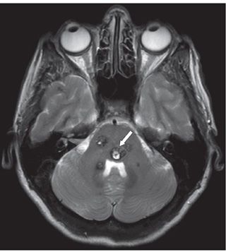
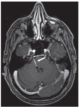
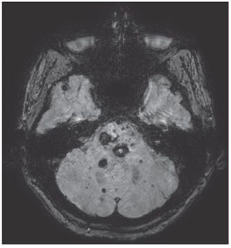
FINDINGS Figure 264-1. Axial T2WI through the pons in patient 1. There is a small focus of susceptibility (signal loss) along the floor of the fourth ventricle on the right (arrow). Figure 264-2. Axial post-contrast T1WI. There is an associated linear enhancing structure (arrow). Figure 264-3. Axial T2WI in patient 2 through the brainstem. There are multiple well-circumscribed lesions with mixed, hyper- and hypointensities without surrounding edema. The dominant lesion (arrow) is T2 hyperintense with a rim of hypointensity and is somewhat bubbly or popcorn-like in morphology. Figure 264-4. Axial GRE through same level. There are many more lesions, 15 to 20 are seen; many of which are punctate both in the brainstem and in the cerebellar hemispheres.


Stay updated, free articles. Join our Telegram channel

Full access? Get Clinical Tree


