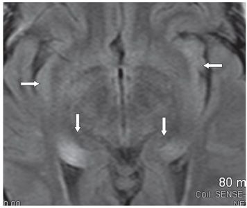
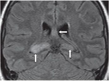
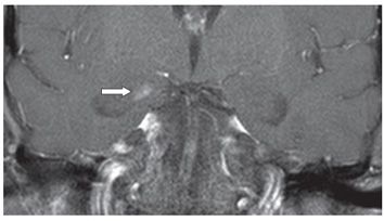
FINDINGS Figure 267-1. Axial MR FLAIR through the temporal lobes. There is bilateral almost symmetrical mesiotemporal smudgy hyperintensity (arrows). Figure 267-2. Axial FLAIR through the hippocampi. There is asymmetric bilateral hippocampal hyperintensity (vertical arrows) and hyperintense insula cortex bilaterally (transverse arrows). Figure 267-3. Coronal FLAIR through hippocampi. There is bilateral asymmetric hippocampal hyperintensity larger on the right than the left (vertical arrows). The transverse arrow points to the hyperintense fornix. Figure 267-4. Coronal post-contrast T1WI through the temporal lobes showing a small right mesiotemporal lobe contrast enhancement (transverse arrow).
DIFFERENTIAL DIAGNOSIS Limbic encephalitis (paraneoplastic), herpes simplex encephalitis (HSE) and human herpes virus 6 (HHV6) encephalitis, seizure-related changes, gliomatosis.
DIAGNOSIS Paraneoplastic limbic encephalitis (PLE).
DISCUSSION


Stay updated, free articles. Join our Telegram channel

Full access? Get Clinical Tree


