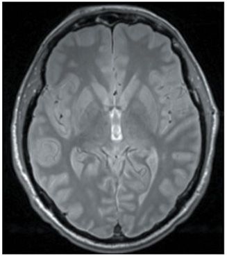
FINDINGS Figure 268-1. This is an abnormal 123I-FP-CIT (DAT scan) SPECT. There is symmetrical decrease of tracer uptake in the bilateral putamen indicating loss of dopamine transporters (DATs) compatible with the diagnosis of Parkinson disease (PD). Figure 268-2. Axial T2 MRI through the basal ganglia. There are no structural abnormalities in the bilateral basal ganglia.
DIFFERENTIAL DIAGNOSIS Normal pressure hydrocephalus (NPH), Parkinson-plus syndromes (progressive supranuclear palsy, multisystem atrophy, corticobasal degeneration), dementia with Lewy bodies, Alzheimer disease.
DIAGNOSIS PD with early cognitive impairment.
DISCUSSION
Stay updated, free articles. Join our Telegram channel

Full access? Get Clinical Tree


