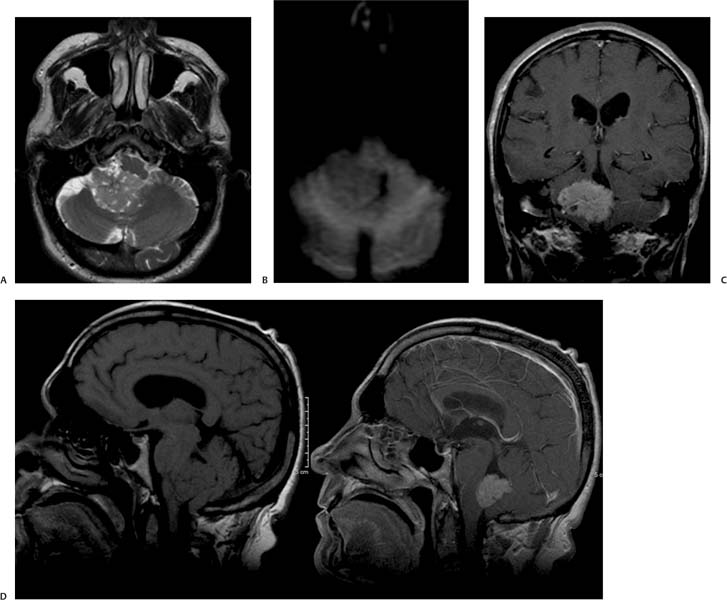
Case 27 A 52-year-old with increasing headaches. (A) Axial T2-weighted image (WI) of the brain shows a heterogeneous mass in the 4th ventricle (arrow) that extends to the right cerebellopontine angle (CPA) through the foramen of Luschka (arrowhead). (B) On axial Diffusion-WI, the mass (arrow) shows no restriction to Diffusion. (C) Coronal T1WI with contrast demonstrates avid enhancement of the 4th ventricle mass and dilatation of the lateral ventricles (arrows). (D) Sagittal T1WIs without and with contrast show a mass with intensity similar to that of the white matter that avidly enhances with gadolinium (arrows). • Choroid plexus papilloma:
Clinical Presentation
Imaging Findings

Differential Diagnosis
![]()
Stay updated, free articles. Join our Telegram channel

Full access? Get Clinical Tree


