 Clinical Presentation
A 65-year-old woman with right hip pain.

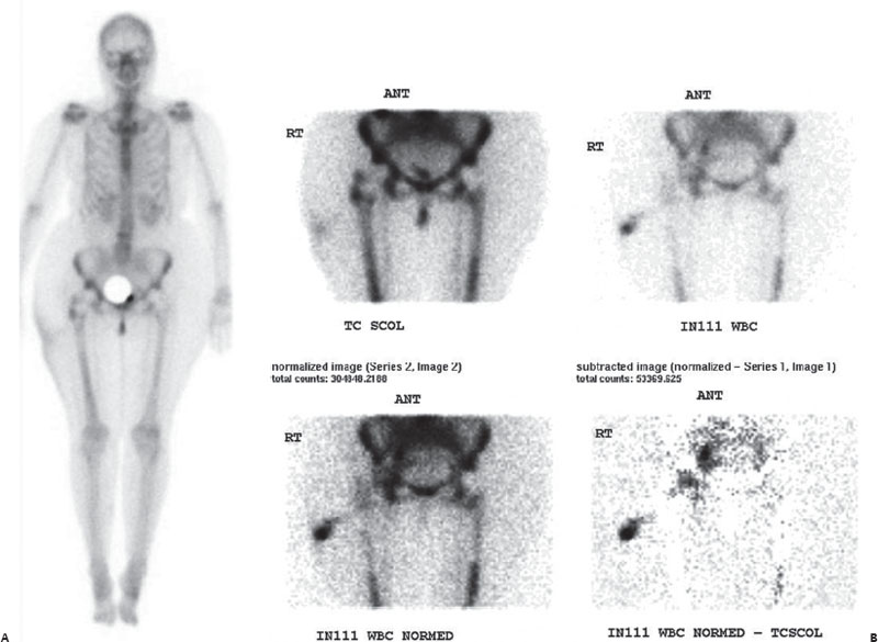
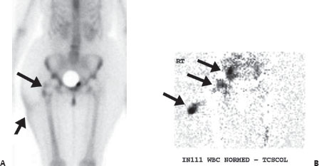
(A) Anterior image from a Tc99m whole-body bone scan reveals increased uptake in the region of the greater trochanter as well as in the region of the femoral neck on the right (long arrow). Photopenic hip hardware is noted bilaterally. Soft-tissue uptake is noted on the lateral aspect of the right upper thigh (short arrow). Mildly increased activity at the right acetabulum is likely degenerative. (B) Simultaneously obtained Tc99m-SCOL and indium 111–tagged WBC scans with different energy windows. Once the two images were obtained, the In111-tagged WBC image was normalized to the Tc99m-SCOL study by counts, and the latter image was subtracted from the former on a pixel-by-pixel basis. The subtracted image reveals discordantly increased In111-tagged WBCs in the lateral thigh soft tissues, right hip joint, and pelvis adjacent to the right hip (arrows).
 Differential Diagnosis
• Infected prosthesis (with right thigh cellulitis and pelvic abscess):
Stay updated, free articles. Join our Telegram channel

Full access? Get Clinical Tree


