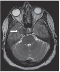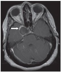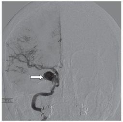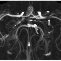


FINDINGS Figure 27-1. Axial NCCT through the sella turcica. There is a well-circumscribed homogeneously hyperdense mass in the right parasellar region (arrow). Figure 27-2. Axial T2WI through the sella. The mass is conspicuously hypointense. Figure 27-3. Axial post-contrast T1WI. There is only a peripheral rim of enhancement (arrow). Figure 27-4. DSA PA right internal carotid injection. The mass opacifies with contrast and arises from the cavernous segment of the right internal carotid artery (ICA) (arrow).
DIFFERENTIAL DIAGNOSIS Aneurysm, cavernous sinus thrombosis, meningioma, metastasis.
Stay updated, free articles. Join our Telegram channel

Full access? Get Clinical Tree


