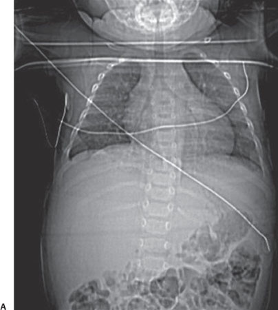
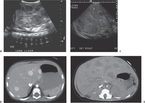
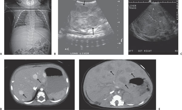
Case 27 A newborn with an abdominal mass. (A) Frontal radiograph of the chest demonstrates cardiomegaly and hepatomegaly. (B,C) Gray-scale and Doppler images of the liver demonstrate multiple hypoechoic lesions (arrows). There is high flow around and within the lesion. (D,E) Early and late post-contrast axial computed tomography (CT) images demonstrate a difference in the caliber of the aorta above and below the celiac artery, as well as multiple hypodense, intensely enhancing lesions in the liver (arrows). • Hemangioendotheliomatosis: These findings are characteristic of hemangioendotheliomatosis. • Metastatic disease:
Clinical Presentation
Further Work-up
Imaging Findings
Differential Diagnosis
![]()
Stay updated, free articles. Join our Telegram channel

Full access? Get Clinical Tree


