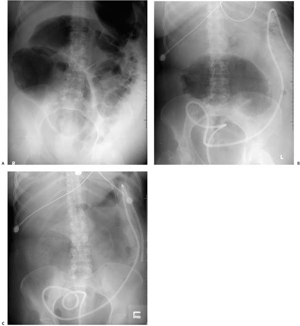
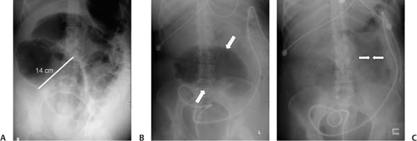
Case 27 A 67-year-old woman presents with abdominal distension and pain. Three serial radiographs were obtained over 2 days. (A) Frontal abdominal radiograph shows a dilated loop of bowel in the right lower quadrant measuring 14 cm in diameter. (B) After placement of a tube through the rectum for decompression, the entire colon decompressed except for this dilated loop (arrows). (C) Follow-up radiograph shows decompression of the dilated loop with massive intra-abdominal free air, as evidenced by the Rigler sign (arrows). • Cecal volvulus with rupture: This is the most likely diagnosis, indicateds by the dilated, air-filled proximal colon (> 10 cm), by failure to evacuate this segment with a rectal drain despite decompression of the transverse and distal colon, as well as by the ultimate finding of pneumoperitoneum. Cecal volvulus more typically presents as a midline or left upper quadrant dilated loop on radiographs, although the appearance can vary based on the length of the mesentery and mobility of the cecum. • Cecal bascule:
 Clinical Presentation
 Imaging Findings
 Differential Diagnosis
![]()
Stay updated, free articles. Join our Telegram channel

Full access? Get Clinical Tree


