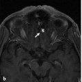
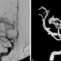
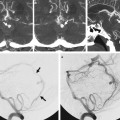
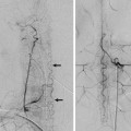
Fig. 27.1 MRA of the neck (a) shows a high-grade stenosis of the proximal intracranial right VA or V4 segment (white arrow). Incidentally, there is a right ECA occlusion (asterisk). Right VA angiogram in anteroposterior (AP) (b) and lateral (c) views and a three-dimensional reconstruction image (d) show contrast filling of both the occipital artery and the APhA through an anastomosis via the artery of the hypoglossal canal (arrows) and musculocutaneous branches from the C1 segment. There is retrograde flow down to the origin of the ECA and opacification of the facial artery (arrowheads).
27.1.3 Diagnosis
Right ECA occlusion with retrograde reconstitution of the ECA through the upper cervical anastomoses from the VA.
27.2 Embryology and Anatomy
The ascending pharyngeal artery (APhA) corresponds to the remnant of the embryonic hypoglossal artery (HA). The HA originates from the third aortic arch and contributes to the development of the most proximal aspect of the cervical ICA. It is one of the embryonic carotid–vertebrobasilar anastomoses, originating from the proximal cervical ICA, slightly distal to the carotid bulb, and enters the posterior cranial fossa via the hypoglossal canal. The relationship between the third aortic arch and the HA can result in various anastomoses between their adult homologs, the ICA, and the APhA, with the most extreme of the spectrum being the persistent HA. See also Case 4.
In the adult, the APhA forms part of the pharyngo-occpital system, a vascular network in the suboccipital region through which the cervical vertebral and carotid arteries are connected by four routes: the occipital, ascending pharyngeal, vertebropharyngeal (C3), and C4-collateral routes.
The APhA originates most commonly from the posterior wall of the proximal ECA and anastomoses with the VA, typically at two levels. The more proximal one occurs laterally between the musculospinal branch of the APhA and the C3 radicular anastomotic branch of the VA. The distal one is the prevertebral branch via the odontoid arch, which usually arises from the HA and follows the same path as the persistent HA, serving as one of the collateral routes in cases of proximal ECA or VA occlusion, as seen in the index case.
Stay updated, free articles. Join our Telegram channel

Full access? Get Clinical Tree


