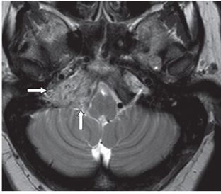
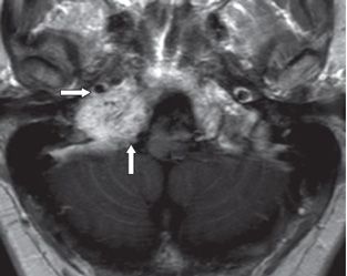
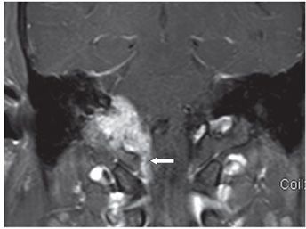
FINDINGS Figures 270-1 and 270-2. Axial FLAIR and T2WI through the jugular foramina. There is a 2.5-cm mass with the epicenter in the right jugular foramen predominantly hyperintense with punctate/linear signal voids (transverse arrows) creating the so-called salt-and-pepper appearance. The margin is irregular. The mass extends to the right CPA (vertical arrows). Figures 270-3 and 270-4. Axial and coronal post-contrast T1WI through the mass. The mass is avidly contrast enhancing within the right jugular foramen. The tumor abuts the internal carotid artery (ICA) (transverse arrow in Figure 270-3) and extends posteriorly to the cerebellopontine angle (CPA) (vertical arrow in Figure 270-3). There is inferior extension into the upper cervical spinal canal (arrow in Figure 270-4).
DIFFERENTIAL DIAGNOSIS Glomus jugulare (GJ), petrous apicitis or osteomyelitis, schwannoma, hypervascular metastasis.
DIAGNOSIS Paraganglioma (GJ).
DISCUSSION


Stay updated, free articles. Join our Telegram channel

Full access? Get Clinical Tree


