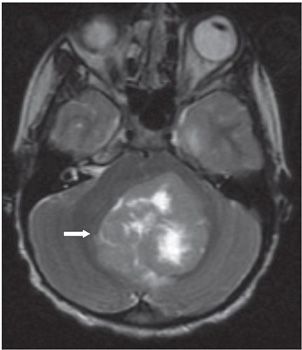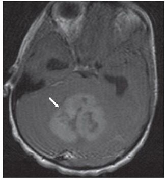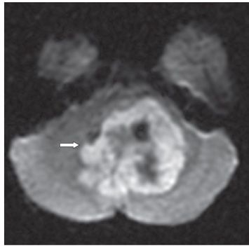


FINDINGS Figure 271-1. Axial NCCT through the posterior fossa. There is a heterogeneous hyperdense mass (white arrow) with central hypodensity indicating intratumoral cysts or necrosis. Expansion of the fourth ventricle around the tumor indicates this is an intraventricular mass. Periventricular hypodensity (black line arrows) reflects transependymal flow of cerebrospinal fluid (CSF) associated with obstructive hydrocephalus. Figure 271-2. Axial T2WI through the mass. The mass is isointense to gray matter (GM) (arrow) with hyperintense intratumoral cysts or necrosis. Figure 271-3. Axial post-contrast T1WI through the mass. There is mild enhancement of the solid component of the mass (arrow). Figure 271-4. Axial DWI through the mass. The mass demonstrates restricted diffusion (arrow).
DIFFERENTIAL DIAGNOSIS Ependymoma, atypical teratoid/rhabdoid tumor (AT/RhT), choroid plexus papilloma, pilocytic astrocytoma, and exophytic brainstem glioma.
DIAGNOSIS Medulloblastoma.
DISCUSSION
Stay updated, free articles. Join our Telegram channel

Full access? Get Clinical Tree


