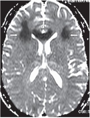
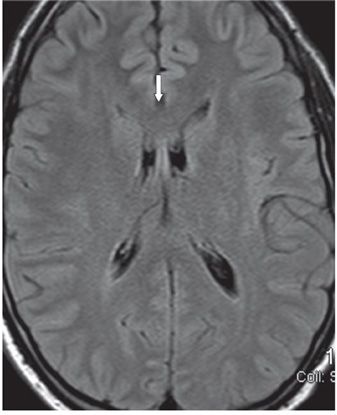
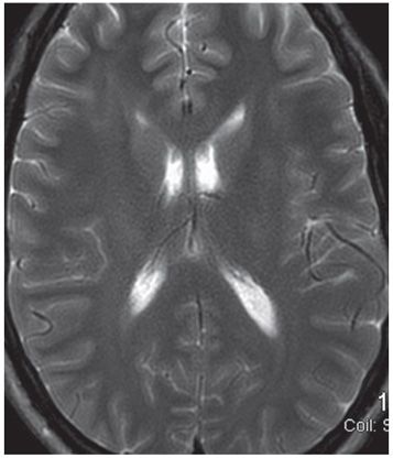
FINDINGS Figures 272-1 and 272-2. Axial DWI with corresponding ADC map through the genu of corpus callosum (CC). There is restricted diffusion in the genu of the CC (vertical arrow) with almost symmetrical restricted diffusion in the bilateral frontal corona radiata (transverse arrows). Figures 272-3 and 272-4. Axial T2 FLAIR and T2WI through the same level show subtle hyperintensity in the CC and white matter (WM)! Other lesions not shown include bilateral multifocal cerebellar T2 hyperintensities. A repeat study 2 days after these images showed that lesions were fading on DWI with isointense areas on FLAIR and T2WI.
DIFFERENTIAL DIAGNOSIS Posterior reversible encephalopathy syndrome (PRES), methotrexate leukoencephalopathy (MTX LE), infarct.
DIAGNOSIS Corpus callosum Methotrexate leukoencephalopathy (MTX LE).
DISCUSSION


Stay updated, free articles. Join our Telegram channel

Full access? Get Clinical Tree


