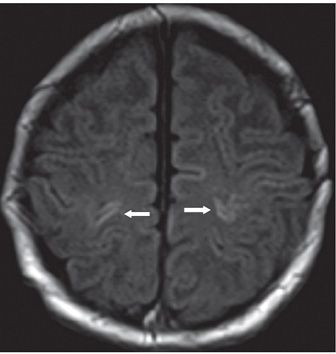
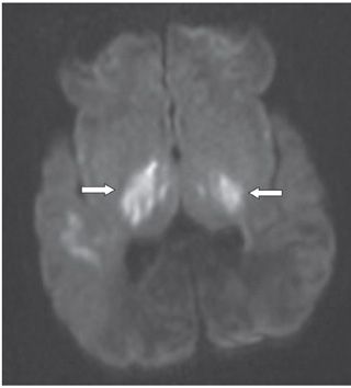
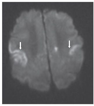
FINDINGS Figure 273-1. Axial non-contrast T1WI through the basal ganglia. There is bilateral symmetrical basal ganglia hyperintensity (arrows) with involvement of the internal capsules and lateral thalami. Figure 273-2. Axial non-contrast T1WI through the high convexities. There is bilateral perirolandic cortical gray matter (GM) hyperintensity (arrows). Figures 273-3 and 273-4. Axial DWI through the basal ganglia and the high convexities, respectively. There is diffusion restriction (arrows) in the bilateral basal ganglia and the perirolandic cortical GM as in Figures 273-1 and 273-2.
DIFFERENTIAL DIAGNOSIS Trauma with diffuse brain swelling, hypoglycemia, maple syrup urine disease, and nonketotic hyperglycinemia (NKHG), hypoxic-ischemic encephalopathy (HIE) in term baby.
DIAGNOSIS HIE in term infants.
DISCUSSION


Stay updated, free articles. Join our Telegram channel

Full access? Get Clinical Tree


