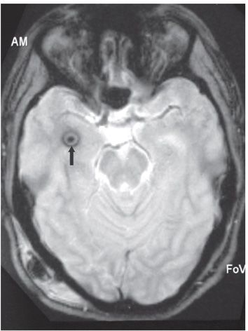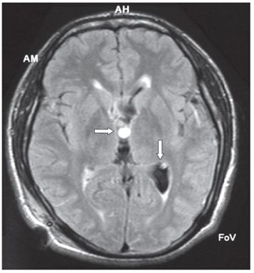

FINDINGS Figure 274-1. Sagittal T1WI. There is a round hyperintense mass within the third ventricle (vertical arrow). Two anterior knobbly components show isointensity and hyperintensity (transverse arrow). Figure 274-2. Axial GRE through the temporal lobes. There is a target hypointense ring in the right temporal horn (arrow). Figure 274-3. Axial FLAIR through the third ventricle. There is a round hyperintensity within the anterior third ventricle (transverse arrow). There is a similar but smaller round hyperintensity in the left trigone (vertical arrow) with asymmetric dilatation of the left trigone.
DIFFERENTIAL DIAGNOSIS Colloid cyst, intraventricular neurocysticercosis (INC), xanthogranuloma.
DIAGNOSIS Intraventricular neurocysticercosis (INC).
DISCUSSION
Stay updated, free articles. Join our Telegram channel

Full access? Get Clinical Tree


