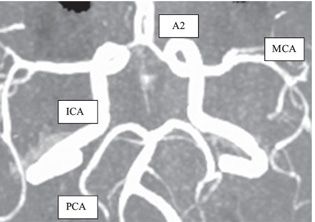
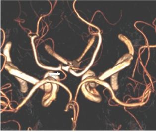
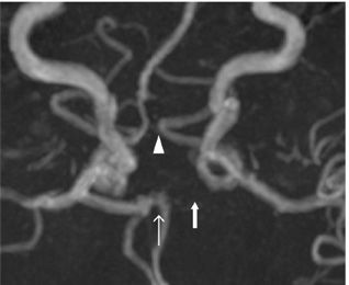
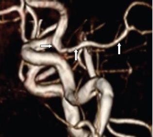
FINDINGS All images are MIP or volume-rendered 3D TOF MRA of the head without contrast rotated to give maximum visualization of the structures intended.
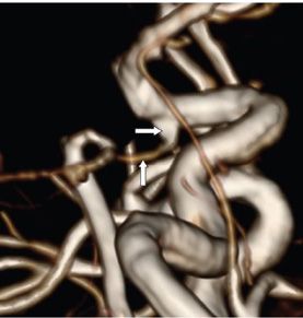
Figure 275-1. Adult-type complete COW. Bilateral A1 (first portion of the anterior cerebral artery [ACA] before the anterior communicating artery [A-Com]—vertical arrows), bilateral asymmetric posterior communicating arteries (P-Com) (transverse arrows), and bilateral P1 (proximal posterior cerebral artery [PCA] from the basilar artery—line arrows) are visualized. The A-Com joins the bilateral A1 in the midline. Figure 275-2. This is the more common pattern where the P-Com is not visualized on the MIP images but visible on the source images (not shown). Figure 275-3. The right P-Com is visualized but not the left on this volume-rendered image. The left P-Com is visible on the source images (not shown). Figure 275-4. The left A1 (arrow points to location) and the left P1 (arrow head points to location) are hypoplastic or absent. The left PCA has a direct origin from the left internal carotid artery (ICA). The A-Com (line arrow) serves as the conduit of blood to the left A2. Figure 275-5


Stay updated, free articles. Join our Telegram channel

Full access? Get Clinical Tree


