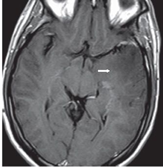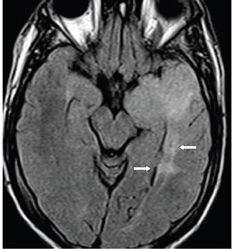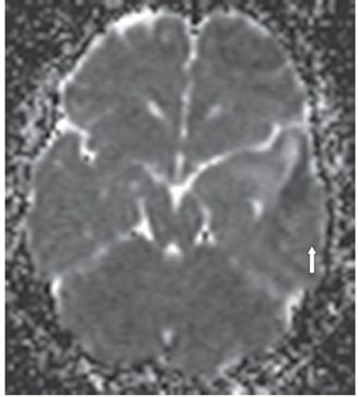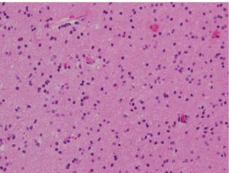


FINDINGS Figure 276-1. Axial non-contrast T1WI through the temporal lobes. There is an expanded left temporal lobe with mild to moderate hypointensity. The abnormality is greater laterally but extends to the medial aspect of the temporal lobe. There is mass effect on the left cerebral peduncle (arrow). Figure 276-2. Axial post-contrast T1WI through same level as Figure 276-1. There is no enhancement. The hypointensity is more profound laterally (arrow) than medially. Figure 276-3. Axial T2 FLAIR. There is diffuse hyperintensity in the expanded left temporal lobe. The abnormality extends more posteriorly along the temporal horn and trigone (arrows). Figure 276-4. Axial ADC map through the mass. There is elevated diffusion in the anterior aspect of the mass with restricted diffusion through the lateral cortex (arrow) with extension into the insula (not shown). Figure 276-5. Photomicrograph shows slightly cellular tumor with round to mildly irregular hyperchromatic nuclei.
DIFFERENTIAL DIAGNOSIS Infarct, herpes encephalitis, astrocytoma WHO II, dysembryoplastic neuroepithelial tumor (DNET), ganglioglioma, pleomorphic xanthoastrocytoma (PXA), seizure-induced abnormality or contusion.
DIAGNOSIS Diffuse astrocytoma WHO II.
DISCUSSION
Stay updated, free articles. Join our Telegram channel

Full access? Get Clinical Tree


