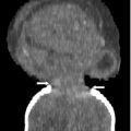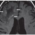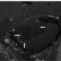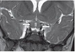
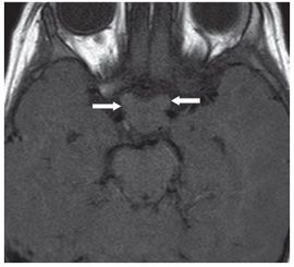
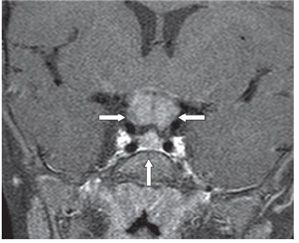
FINDINGS Figure 277-1. Axial FLAIR MRI through the suprasellar region. There is a 1.3 cm × 2 cm mildly lobulated isointense mass in the region of the chiasm (arrows). Figure 277-2. Coronal T2WI slightly anteriorly to the chiasm. There is thickening of bilateral isointense (to gray matter [GM]) optic nerves (arrows). Figure 277-3. Axial T1WI through the suprasellar cistern. There is an isointense mass in the chiasm with bilateral posterior optic nerves thickening (arrows). Figure 277-4. Coronal post-contrast T1WI through the mass. There is homogeneous contrast enhancement of the mass (transverse arrows). The mass is clearly separated from the pituitary gland (vertical arrow).
DIFFERENTIAL DIAGNOSIS Optic pathway glioma (OPG), lymphoma, metastasis, sarcoidosis, granuloma, craniopharyngioma.
DIAGNOSIS Optic pathway glioma (OPG).
DISCUSSION
Stay updated, free articles. Join our Telegram channel

Full access? Get Clinical Tree


