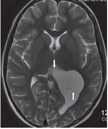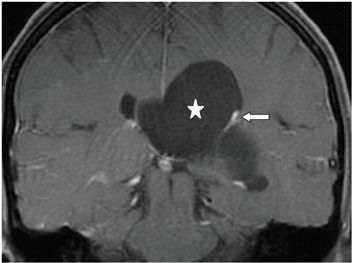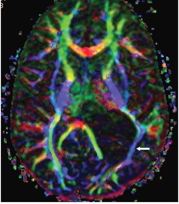


FINDINGS Figure 278-1. Axial FLAIR through the lateral ventricles. There is distension of the left lateral ventricle by a large CSF intensity cyst (star) which displaces the septum pellucidum medially (transverse arrow) and the left choroid plexus posterolaterally (vertical arrow). It is difficult to make out the cyst wall except where it compresses the ventricular walls laterally and medially. Figure 278-2. Axial T2WI through the trigones. The cyst is of CSF intensity with a very thin dark membranous wall (arrows). There is dilatation of the left occipital horn. Figure 278-3. Coronal post-contrast T1WI. The outline of the cyst (star) blends with the compressed lateral ventricular wall. The displaced enhancing left choroid plexus is compressed against the lateral ventricular wall (arrow). Figure 278-4
Stay updated, free articles. Join our Telegram channel

Full access? Get Clinical Tree


