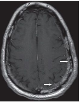
FINDINGS Figure 279-1. Axial NCCT through the cranial vault bone window. There is a wide region of mottled hypodensity in the left parietal bone with smaller similar areas elsewhere (arrows). Figure 279-2. Axial post-contrast T1WI through the same region. There is enhancement within the diploic space which correlates with hypodensities on CT. There is perhaps underlying epidural thickening (arrows).
DIFFERENTIAL DIAGNOSIS Multiple myeloma, metastases, osteomyelitis, eosinophilic granuloma.
DIAGNOSIS Osteomyelitis.
DISCUSSION
Stay updated, free articles. Join our Telegram channel

Full access? Get Clinical Tree


