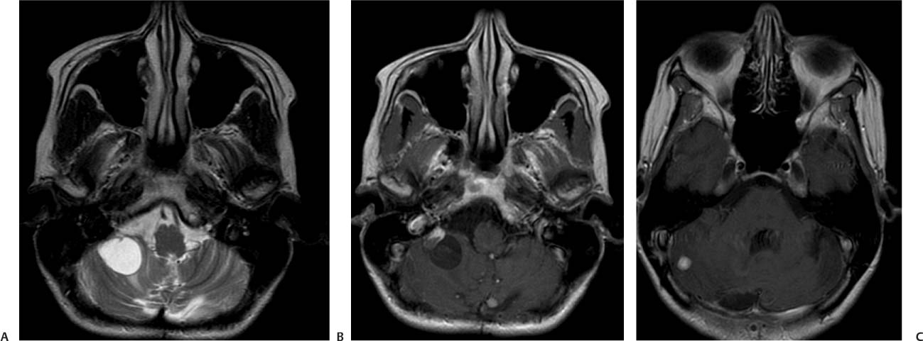
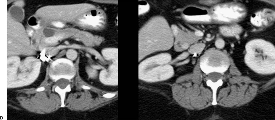
Case 28 A 54-year-old woman with a recent history of vertigo. (A) Axial T2-weighted image (WI) of the posterior fossa shows a cystic lesion on the right cerebellar hemisphere (arrow). There is surrounding vasogenicedema. (B) On axial T1WI with contrast, the right cerebellar lesion shows a mural nodule (white arrow); the cystic component (black arrow) does not enhance. A second enhancing lesion (arrowhead) is noted near the midline. (C) Axial T1WI of the posterior fossa with contrast shows another enhancing lesion on the left cerebellar hemisphere (arrow). (D) Axial computed tomography (CT) scans of the abdomen with contrast show a simple pancreatic cyst (white arrow). A small enhancing lesion on the pancreatic head is also seen (black arrow).
Clinical Presentation
Further Work-up
Imaging Findings
Stay updated, free articles. Join our Telegram channel

Full access? Get Clinical Tree


