 Clinical Presentation
A 55-year-old man with progressive shortness of breath who has abnormal findings on chest radiography.
 Imaging Findings
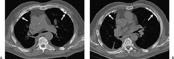
(A, B) Non–contrast-enhanced computed tomography (CT) of the chest shows pleural thickening and extensively calcified pleural plaques bilaterally (arrows).
 Differential Diagnosis
• Asbestos-related pleural plaques: Bilateral calcified pleural plaques are a characteristic presentation of asbestos-related pleural disease.
• Postinfectious pleural plaques:
Stay updated, free articles. Join our Telegram channel

Full access? Get Clinical Tree


