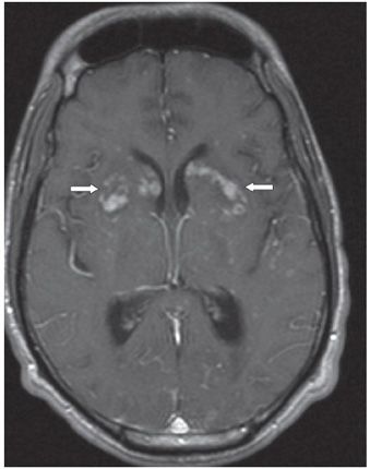
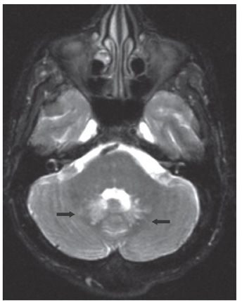
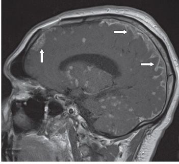
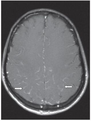
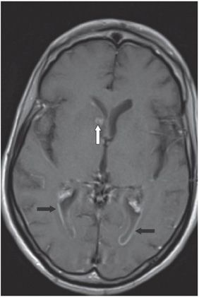
FINDINGS Figure 28-1. Axial FLAIR through the basal ganglia. There is bilateral symmetrical basal ganglia hyperintensity (arrows). Mild hydrocephalus is present along with other parenchymal lesions. Figure 28-2. Corresponding post-contrast T1WI. There are nodular and ring enhancement in the basal ganglia bilaterally (arrows) with smaller ones elsewhere in the cerebral hemispheres. Figure 28-3. In the same patient, axial T2WI through the posterior fossa shows dilated and hyperintense perivascular spaces in the region of the dentate nuclei (arrows) of the cerebellum, suggesting cryptococcal involvement. Figure 28-4. Parasagittal post-contrast T1WI in a different patient shows multiple brain parenchyma-enhancing nodules (including the cerebellum) and thick leptomeningeal enhancement (arrows). Figure 28-5. Axial post-contrast T1WI (companion patient) through the centrum semiovale shows thin leptomeningeal enhancement (arrows). Figure 28-6. Axial post-contrast T1WI shows ependymal enhancement (arrows) in the trigones of the lateral ventricles and a nodular lesion in the head of the right caudate nucleus (vertical arrow).
DIFFERENTIAL DIAGNOSIS
Stay updated, free articles. Join our Telegram channel

Full access? Get Clinical Tree


