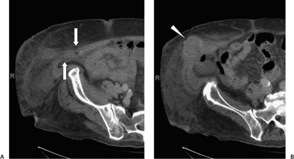
Case 28 A 65-year-old man presents with a palpable mass in the right lower quadrant of the abdomen. (A) Linear strands of soft tissue suggestive of mesenteric blood vessels cross a defect in the abdominal wall just anterior to the iliac spine, between the fibers of the rectus abdominis muscle and the fibers of the transversalis abdominis and internal oblique muscles (arrows). (B) A pocket of gas lies just at the abdominal wall defect within the abdomen, and a thick-walled, fluid-filled structure (arrowhead) lies peripheral to this gas pocket within the abdominal wall. • Spigelian hernia (SH): This is the most likely diagnosis, given the apparent passage of abdominal contents lateral to the rectus abdominis muscle through the transversalis abdominis and internal oblique muscles. • Abdominal wall abscess:
 Clinical Presentation
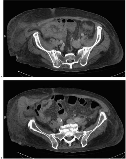
 Imaging Findings
 Differential Diagnosis
![]()
Stay updated, free articles. Join our Telegram channel

Full access? Get Clinical Tree


