The Cranial Nerve Supply
28.1 Case Description
28.1.1 Clinical Presentation
An 82-year-old, right-handed woman presented with a several-month history of falls and gait ataxia, worse on the left side, which prompted further imaging.
28.1.2 Radiologic Studies
See ▶ Fig. 28.1, ▶ Fig. 28.2.
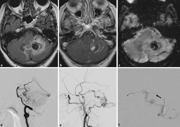
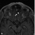
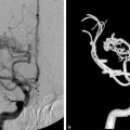
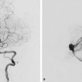
Fig. 28.1 MRI axial fluid-attenuated inversion recovery (a), T1 contrast-enhanced (b), and susceptibility (c) weighted images demonstrate a vascular pouch in the left inferior cerebellum with significant perifocal edema, as well as abnormal sulcal vessels. Left vertebral artery angiogram in lateral view (d) reveals a cerebellar arteriovenous malformation, the imaging characteristics of which cannot explain the visualized pouch on MR. Subsequent injection into the left ECA (e) demonstrates a dural arteriovenous fistula along the petrous ridge, with arterial filling via the meningohypophyseal trunk and the facial arcade and multiple venous pouches and venous stenoses. After embolization of the distal MMA with glue deposition into the venous segment, minimal residual flow was visualized via the petrosal branch of the MMA (injection in f). From this vessel, transient opacification of a normal distal branch was seen that connected to the stylomastoid branch of the posterior auricular artery (arrow). As glue had been deposited in the foot of the vein, we only ligated the petrosal branch of the MMA with concentrated glue to ensure that the supply to the facial nerve was not compromised and the flow to the fistula was further reduced, which was supposed to facilitate further thrombosis. Case continued in ▶ Fig. 28.2.
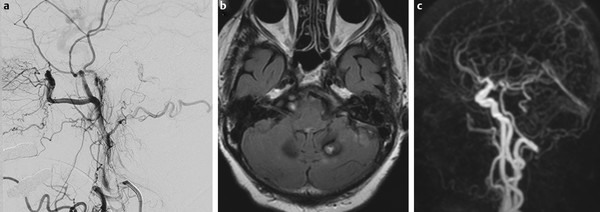
Fig. 28.2 Immediate control angiogram (a) demonstrated residual very slow filling of the shunt via the stylomastoid branch of the posterior auricular artery. Upon follow-up MR 2 weeks later (b), the patient had thrombosed her venous pouch, the edema was no longer visualized, and only the arteriovenous shunting related to the cerebellar arteriovenous malformation was noted on the dynamic contrast-enhanced magnetic resonance angiography, with no further shunting visualized from the dural branches. (c) Clinically, she did very well, with no new neurological deficits and reversal of her presenting symptoms.
28.1.3 Diagnosis
Dural arteriovenous fistula (dAVF) of the petrous ridge with cortical venous reflux. The dAVF is fed by the middle meningeal artery (MMA), including its petrosal branch and the stylomastoid branch of the posterior auricular artery (i.e., the facial arcade).
28.2 Anatomy
Under normal conditions, the caliber of the arteries that supply the cranial nerves (the vasa nervorum) is between 100 and 300 μm, and they are, therefore, in most instances only visible on conventional angiography during superselective injections. Nevertheless, knowledge of their origin, their anastomoses, and their potential variations is paramount for safe embolization of a variety of skull base lesions, including dAVF and hypervascularized tumors.
28.2.1 Cranial Nerves I and II
Being outpouchings of the brain, rather than true cranial nerves, the olfactory and optic nerves are not considered peripheral nerves and will be described only briefly: the olfactory nerve is supplied by the olfactory artery and ethmoidal branches of the ophthalmic artery, and the optic nerve is supplied by the proximal ophthalmic artery.
28.2.2 Cranial Nerves III, IV, and VI
In its cisternal segment, the oculomotor nerve receives arterial supply in the vicinity of the posterior perforating substance from the basilar or the posterior cerebral artery.
Once it extends forward from the brainstem, the trochlear nerve is supplied by the superior cerebellar artery and additional circumferential arteries of the P1 segment of the posterior cerebral artery, with which it courses through the ambient cistern.
The abducens nerve is supplied by the clival dural network along its course anterior to the pons, including the medial and lateral clival arteries from the hypoglossal and jugular branch of the neuromeningeal trunk of the ascending pharyngeal artery and, more cranially, the medial and lateral clival arteries from the meningohypophyseal trunk.
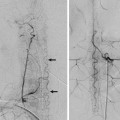
These dural arteries also contribute to the supply of cranial nerves III, IV, and V in their dural and transosseous course. The third and fourth cranial nerves are supplied by the artery of the free margin of the tentorium on the roof of the cavernous sinus. This artery may arise from the meningohypophyseal trunk, directly off the internal carotid artery (ICA), but also from the MMA, the ophthalmic or lacrimal artery, or the inferolateral trunk (ILT). Further distally (i.e., in the cavernous sinus and the superior orbital fissure), the anteromedial branch of the ILT supplies nerves III, IV, and VI (▶ Fig. 28.3).
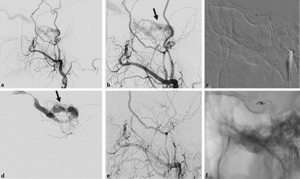
Fig. 28.3 This patient had a dural arteriovenous fistula of the cavernous sinus that was supplied by multiple interconnecting internal maxillary branches that all converged into a single artery (arrows
Stay updated, free articles. Join our Telegram channel

Full access? Get Clinical Tree


