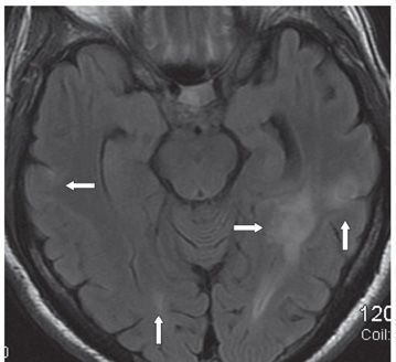
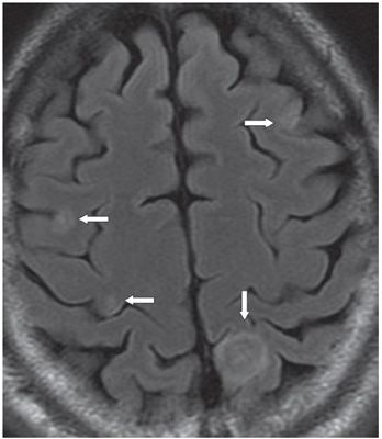
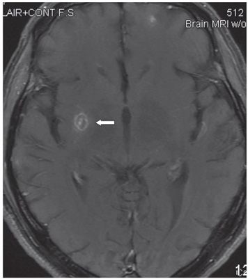
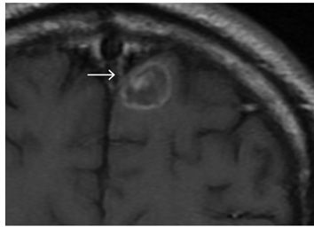
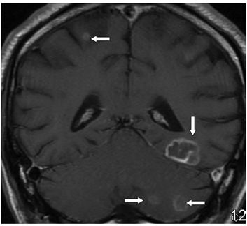
FINDINGS Figure 281-1. Axial non-contrast T1WI through the vertex. There is a left parietal parafalcine cortical somewhat isointense (to surrounding gray matter) round lesion (arrow). There is a central linear cleft and a thin peripheral hyperintensity. Figure 281-2. Axial MR FLAIR through the temporal lobes. There are multiple lesions in bilateral temporal lobes and right occipital lobe (arrows). Within what appears to represent smudgy hyperintensities (edema) are well-defined isointense round lesions. Figure 281-3. Axial FLAIR through the left parietal lesion. The mass shows alternating rings of hyperintensity and isointensity (concentric target sign) also demonstrated on T2WI (not shown) (vertical arrow). There are additional smaller cortical lesions in bilateral frontal cortex (transverse arrows) showing similar concentric target sign. Figure 281-4. Axial post-contrast T1WI through the basal ganglia. There is a thin ring-enhancing lesion in the right lentiform nucleus with a central contrast-enhancing nodule (arrow). Figures 281-5 and 281-6
Stay updated, free articles. Join our Telegram channel

Full access? Get Clinical Tree


