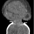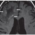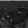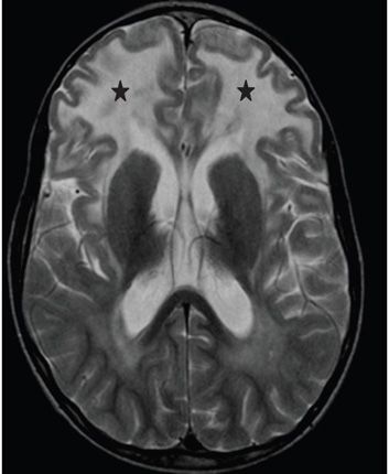
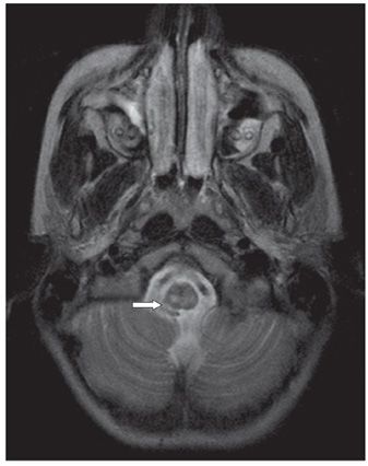
FINDINGS Figure 282-1. Axial T2WI through the centrum semiovale. There is extensive confluent frontal and parietal white matter (WM) hyperintensity involving deep and subcortical regions (stars). Figure 282-2. Axial T2WI at the level of the lateral ventricles. There is predominant bilateral frontal lobe WM confluent hyperintensity (stars). Figure 282-3. Axial T2WI through the medulla. Areas of hyperintensity (arrow) are seen in the upper medulla too. (Case courtesy of Dr. Karuna Shekdar, University of Pennsylvania, Philadelphia.)
DIFFERENTIAL DIAGNOSIS Canavan disease, megalencephalic leukoencephalopathy with subcortical cysts (MLC), mucopolysaccharidoses (MPS), Alexander disease.
DIAGNOSIS Alexander disease.
DISCUSSION
Stay updated, free articles. Join our Telegram channel

Full access? Get Clinical Tree


