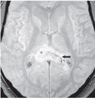
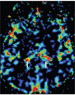
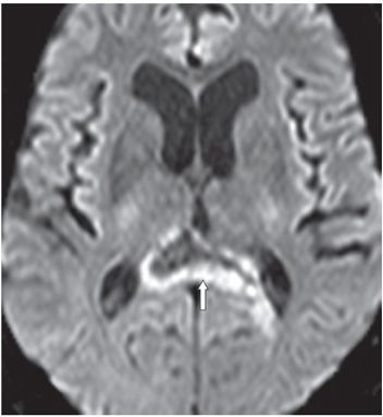
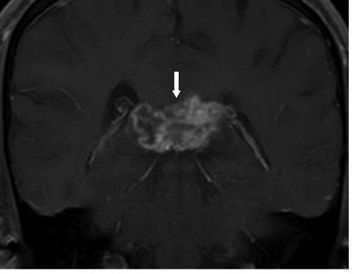
FINDINGS Figure 283-1. Axial FLAIR through the splenium of corpus callosum. There is hyperintense enlargement of the splenium of corpus callosum (CC) (arrow). There is a thick hyperintense rim surrounding a less hyperintense core. Figure 283-2. Axial GRE through same level as Figure 283-1. There is irregular hypointensity within the splenium mass (black arrow) suggestive of hemorrhagic product or calcifications. Figure 283-3. Axial relative Cerebral Blood Volume (rCBV) map. There is relative increased blood volume within the splenium mass (arrows). Figure 283-4. Axial DWI through the splenium. There is peripheral restricted diffusion in the mass suggesting a highly cellular mass. Figure 283-5. Coronal post-contrast T1WI. There is heterogeneous irregular contrast enhancement (arrow).
DIFFERENTIAL DIAGNOSIS Glioblastoma (GB), lymphoma, demyelinating lesion.


Stay updated, free articles. Join our Telegram channel

Full access? Get Clinical Tree


