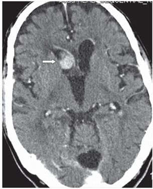
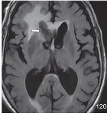
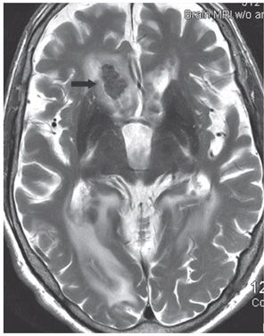
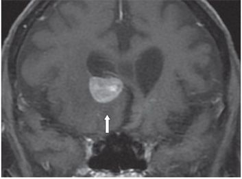
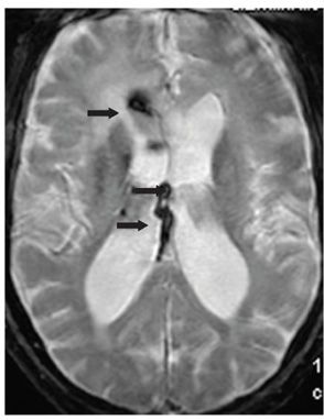
FINDINGS Figures 284-1 and 284-2. Axial NCCT and post-contrast CT, respectively, through the frontal horns. There is an ovoid contrast-enhancing mildly hyperdense mass in the right frontal horn (transverse arrow) with adjacent right frontal white matter (WM) hypodensity (vertical arrow) consistent with edema. There is associated ependymal enhancement. Figure 284-3. Axial FLAIR through the frontal horns. There is an ovoid irregular hypointense mass occupying the right frontal horn (transverse arrow) with surrounding parenchymal hyperintensity reminiscent of vasogenic edema. Figure 284-4. Axial T2WI through the frontal horns. The mass is relatively hypointense to gray matter (GM) (arrow). There is bilateral peritrigonal hyperintensity. Figure 284-5. Coronal post-contrast T1WI through the frontal horns. The right frontal horn mass is avidly contrast enhancing (arrow) and has a smooth margin. Non-contrast T1WI (not shown) demonstrated an isointense mass. Figure 284-6


Stay updated, free articles. Join our Telegram channel

Full access? Get Clinical Tree


