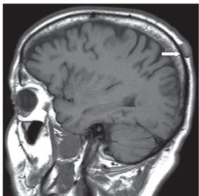
FINDINGS Figure 285-1. Axial T2WI through the parietal bones. There is an ovoid hypointense posterior right parietal scalp nodule (arrow). Figure 285-2. Right parasagittal T1WI through the mass. The nodule is of intermediate intensity almost isointense with brain.
DIFFERENTIAL DIAGNOSIS Dermoid, epidermoid, sebaceous cyst, lipoma.
DIAGNOSIS Sebaceous (trichilemmal or pilar) cyst.
DISCUSSION


Stay updated, free articles. Join our Telegram channel

Full access? Get Clinical Tree


